View over over 4000 high resolution images of histological structures accompanied by interactive descriptive text that labels relevant histological details of every cell and tissue in the human bodyThe central vein is a small vein structurally characterised by its central position in the lobule of the liver functionally characterised by contribution to the larger whole but being the origin of the hepatic venous system that transports metabolically rich products to the rest of the body part of hepatic venous system made up from terminal branches of the sinusoids common diseasesHistology Photomicrographs Human Anatomy and Physiology (BIOL& 241L242L) Karen Hart, Peninsula College Epithelial tissue ;
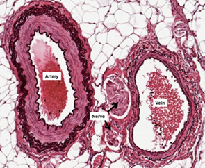
Human Structure Virtual Microscopy
Artery vein nerve histology
Artery vein nerve histology-In anatomical sources, the aorta is usually divided into sections One way of classifying a part of the aorta is by anatomical compartment, where the thoracic aorta (or thoracic portion of the aorta) runs from the heart to the diaphragmThe aorta then continues downward as the abdominal aorta (or abdominal portion of the aorta) from the diaphragm to the aortic bifurcationIn the vein, the same 3 tunics can be seen but the tunica media is reduced, and the tunica adventitia is wider compared to the artery


11 Circulatory System Ideas Circulatory System Arteries And Veins Anatomy And Physiology
Skeletal muscle, smooth muscle;Explore Magaly Chavez's board "artery vein nerve" on See more ideas about arteries, medicine studies, anatomy and physiologyWe've gathered our favorite ideas for Artery And Vein Histology Slide, Explore our list of popular images of Artery And Vein Histology Slide and Download Photos Collection with high resolution
15/7/17 Artery and vein Stain Ladewig's trichrome Low magnification Histological layers Tunica intima With haematoxylin and eosin stains, blood vessels can be easily observed on light microscopy There are three distinct layers forming the walls of arteries and veins The innermost layer is the tunica intimaThe exact anatomy of the central retinal vein as it exits the eye is unknown In this study serial sections of the central retinal vein and artery in the anterior optic nerve from six globes (five from cornea donors and one exenteration specimen) were examined by image analysis and threedimensional reconstruction to determine their luminal characteristicsThis is an online quiz called Artery and nerve histology There is a printable worksheet available for download here so you can take the quiz with pen and paper Your Skills & Rank Total Points 0 Get started!
The layers of the heart conform to the basic pattern seen in the histology of other tubular structure except that the muscle layer dominates Tubular structures in the body have a basic structural makeup of an inner layer lined with an epithelial layer abutting the lumen, a middle functional layer and an outer protective layer or skin/8/12 Vein Histology in Histology Vein has endothelium and a thin subendothelial layer with smooth muscle cells among the connective tissue elements Internal elastic membrane may or may not be present The tunica media is much thinner The tunica adventitia is usually thicker than the media and is mostly made of collagen fibresNerve Endings* Popliteal Artery/anatomy & histology* Popliteal Vein


Human Structure Virtual Microscopy


Histology Page
Presence of terminal nerve corpuscles in the walls of the popliteal artery and vein and in immediate proximity to those vessels and their collaterals Article in Undetermined Language BERTELLI L PMID PubMed indexed for MEDLINE MeSH Terms Arteries* Humans;Blood vessels Light micrograph of a section through tissue showing an artery (middle ) and a vein (top left) Surrounding the artery and vein are layers of smooth muscle (pink), and fibrous connective tissue (orange) Arteries carry blood away from the h https//wwwalamycom/licensesandpricing/?v=1 https//wwwalamy8/2/09 Vascular Artery & Vein Artery Vein 31 Vascular Artery & Vein Artery = Thick walled Vein = Thin walled Connective Tissue (some Epithelial present) Part of the Circulatory system 32 Vascular Artery, Vein & Nerve Nerve Artery Vein 33 Vascular Artery, Vein & Nerve Epithelial Tissue Part of the Circulatory system 34
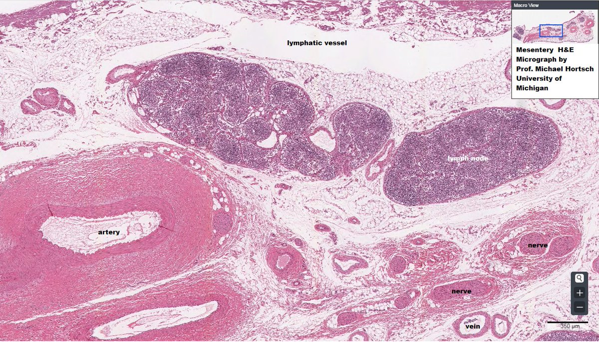


Dr Amanda J Meyer Pronounced M R Easy To Appreciate A Neurovascular Bundle In This Micrograph Artery Vein Lymph Nerve Anatomy Histology Vmdatabase T Co Lo8vq8dfjq


Histology Review Nervous Tissue Dr Tim Ballard Department
The exact anatomy of the central retinal vein as it exits the eye is unknown In this study serial sections of the central retinal vein and artery in the anterior optic nerve from six globes (five from cornea donors and one exenteration specimen) were examined byPrepared slide Cross section of nonhuman artery, vein and nerveArtery and vein Photo about tissue, nerve, micrograph, artery, vascular, system, microscope, vessel, vein, microscopy, adipose, muscular, histology, circulatory
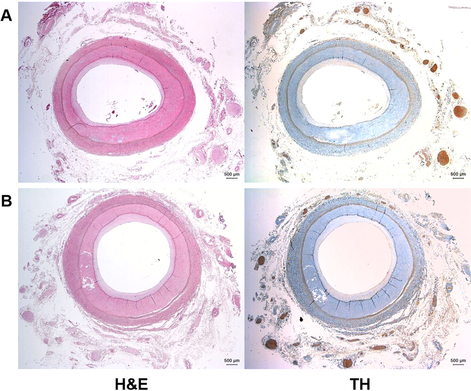


Anatomic Conformation Of Renal Sympathetic Nerve Fibers In Living Human Tissues Scientific Reports


Histology Of Blood Vessels Ppt Video Online Download
Medium sized (muscular) artery and vein (2 virtual slides) Distinct layers of wall of an artery tunica intima, internal elastic lamina, tunica media (many layers of smooth muscle), external elastic lamina (multiple layers) extending into tunica adventitia;Small blood vessels Lymphatic system ;Elastic arteries are primarily represented by the large vessels emerging from the heart ventricles, such as the aorta and the pulmonary artery Characteristic for these vessels are numerous concentric elastic lamellae of the tunica media interspersed with bundles of smooth muscle cells Elastin can be stained with special stains
:background_color(FFFFFF):format(jpeg)/images/library/3598/6B12iEgxEPjcsiKAanmfQ_Tunica_intima.png)


Blood Vessels Histology And Clinical Aspects Kenhub
:background_color(FFFFFF):format(jpeg)/images/library/3613/OgR38mkJaHfLSb8S17ENg_Lymphatic_vessel_of_the_dermis.png)


Blood Vessels Histology And Clinical Aspects Kenhub
Histology This section of dentaljuce has over 400 histological slides, showing tissues from all organ systems in their healthy state Each tissue/organ slide set has an explanatory accompanying text which desribes its structure, function and roleMeyer's Histology Online Interactive Atlas and Virtual Microscope Improve your identification and understanding of histological structures!The parotid gland is a major salivary gland in many animals In humans, the two parotid glands are present on either side of the mouth and in front of both earsThey are the largest of the salivary glands Each parotid is wrapped around the mandibular ramus, and secretes serous saliva through the parotid duct into the mouth, to facilitate mastication and swallowing and to begin the
:background_color(FFFFFF):format(jpeg)/images/library/3610/5NHWYTWfF0zpaLrwavgwQ_Venule.png)


Blood Vessels Histology And Clinical Aspects Kenhub


Cardiovascular
The circulation of the posterior ciliary artery is the main source of blood supply to the ONH, except for the surface nerve fiber layer which is supplied by the retinal circulation The blood supply in the ONH is sectoral in nature, which is the reason why there is the sectoral involvement of the ONH in its ischemic disordersArtery and vein histology 3p Image Quiz PurposeGames Create Play Learn23/9/19 Histology Of Arteries Veins And Capillaries Preview Blood Vessels Lab Blue Histology Vascular System Artery Vein And Nerve Slide Peripheral Arteries Veins And Artery Blood Vessel Under Microscope View For Education Histology Top 60
:background_color(FFFFFF):format(jpeg)/images/library/13219/arteries-and-veins.psb_english.jpg)


Blood Vessels Histology And Clinical Aspects Kenhub


Histology Of Blood Vessels Radiology Reference Article Radiopaedia Org
Vascular Supply of the Kidney Renal Artery, Vein and Nerves Renal Artery The kidneys are supplied by parietal branches of the aorta (renal artery)The vascular supply (A renalis dexter et sinistra) is often subject to variationsThe right renal artery passes under the vena cava toHistology The Common Vein Blood Supply Venous Drainage Lymphatics Nerves Portal vein liver hepatic cirrhosis fx fibrosis fx nodules fx bridging between the triads portal triad hepatic artery portal vein bile duct histopathology Courtesy Barbara BannerYou will be examining a cross section of the artery vein and nerve Histology from BIOLOGY 105 at Queens College, CUNY
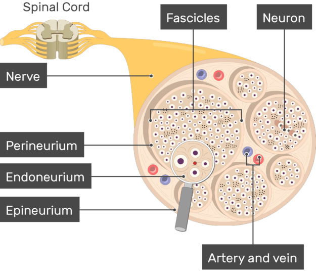


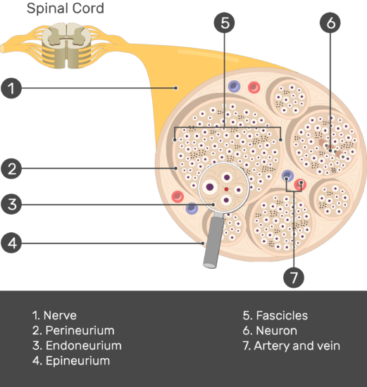
Nerve Structure Anatomy


Untitled Document
Shotgun Histology Medium Artery and Vein6/1/16 Identification points Innumerable transverse sections of myelinated (oligodendrocytes) axons of ganglion cells arranged in fascicles are seen In the middle central retinal artery and central retinal vein is seen At the periphery the nerve is surrounded by three layers of meninges duramater, arachnoidmater and piamater are seenSection, trichrome stain A 10% discount applies if you order more than 10 of this item and 15% discount applies if you order more than 25 of this item Triarch Incorporated offers superior prepared microscope slides


Artery And Vein Digital Slide Youtube
:background_color(FFFFFF):format(jpeg)/images/library/3615/NOpbXd2vHcY0t8xD0mZLQ_Vasa_vasorum.png)


Blood Vessels Histology And Clinical Aspects Kenhub
Single, prepared slide with a cross section of artery, vein & nerve Great tool to study histology of these tissues and compare and contrast their characteristics Slide measures 75mm wide and 25mm long Arrives in a protective cardboard casing Single, prepared microscope slide with a cross section of artery, vein &amSlides are expertly prepared, and labeled for easy identification Single, prepared microscope slide with a cross section of artery, vein & nerve Great tool to study histology of these tissues and compare and contrast their characteristics27/7/21 Arteries, veins and nerves of the trunk (diagram) Arteries of the trunk include the thoracic aorta, celiac trunk, superior mesenteric artery, inferior mesenteric artery, and common iliac arteries (with its terminal branches internal iliac and external iliac arteries )


11 Circulatory System Ideas Circulatory System Arteries And Veins Anatomy And Physiology
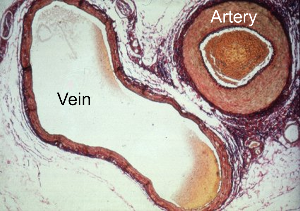


Cardiovascular System
Histology Slides Clemens BIOL 218 Histology Slide Images Exercise 2 Cells 02_Blood_100X 02_Blood_400X 02_Neurons_40X 13_Artery Vein Nerve_40X 13_Artery Nerve_100X 13_Aorta media externa_40X 13_Basophil_400X Muscular Artery Vein And Nerve Bundles Surrounded By Adipose Histology Laboratory Manual Hm Practical Blood Vessel Histology Embryology Vessels Vein And Artery2png Jpg 717 3 Anatomy How To Differentiate A Vein From An Artery Quora Cardiac Muscle Ppt Video Online DownloadMediumsized artery and vein and nerves ts microscope slide In stock
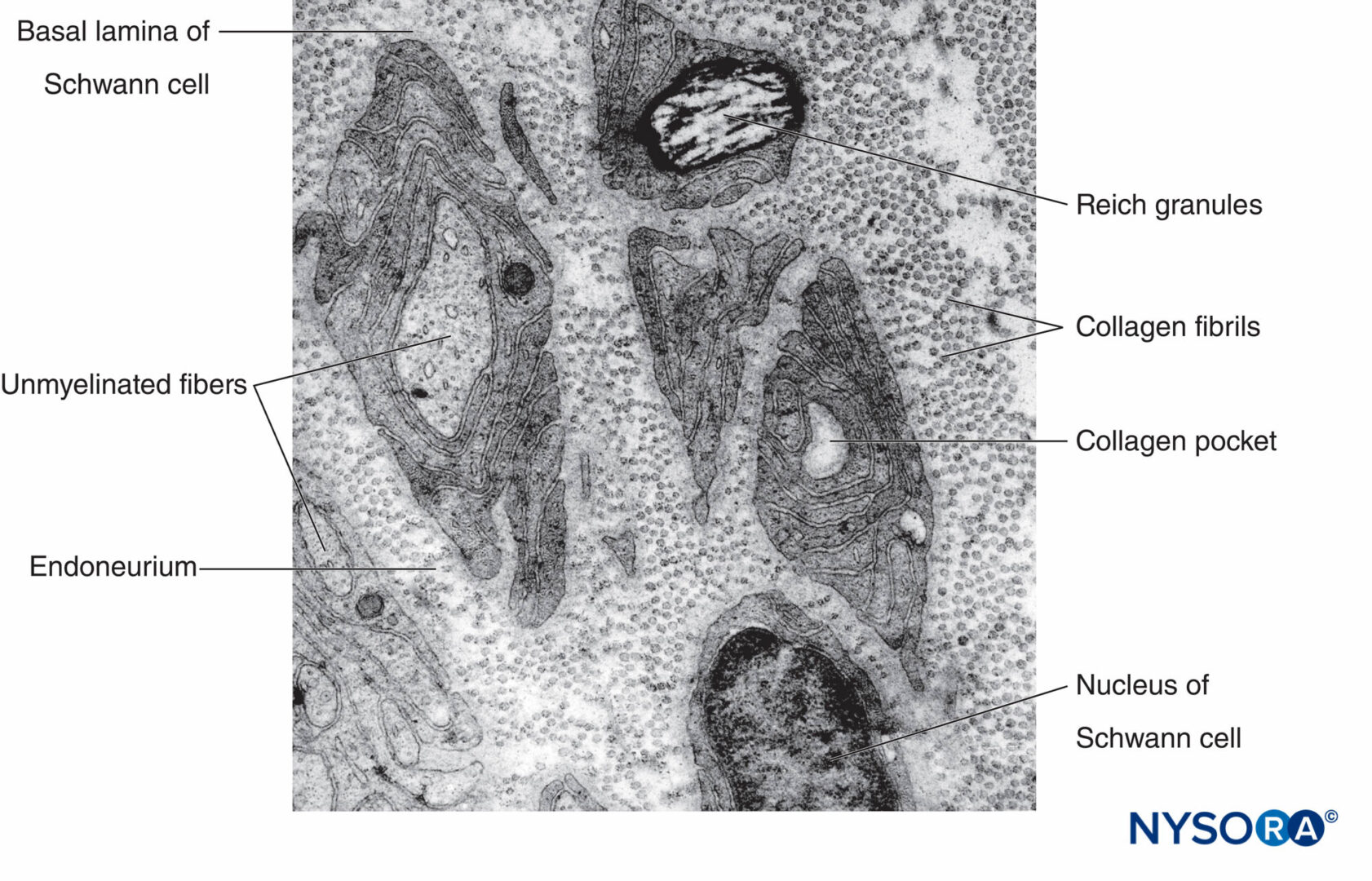


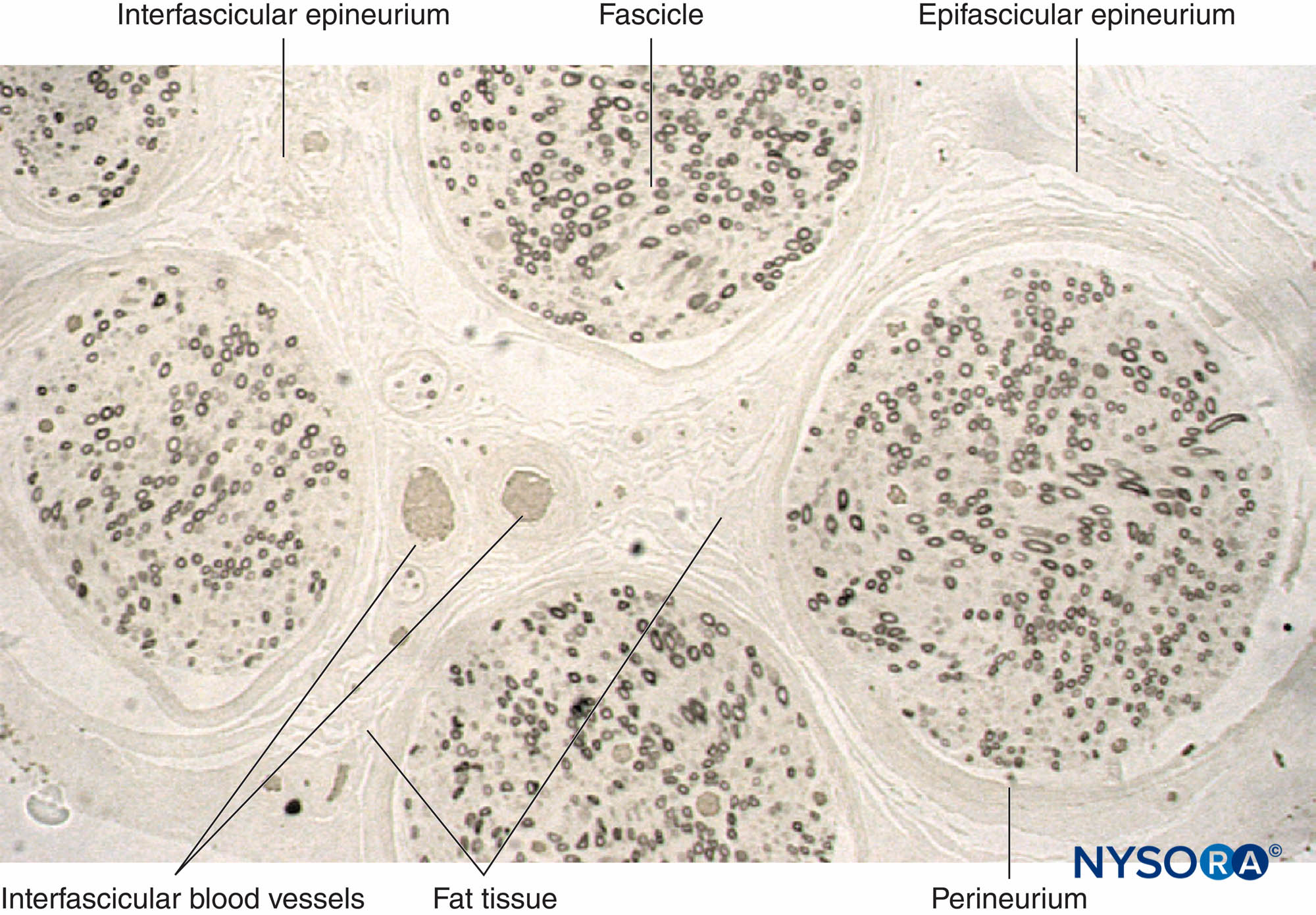
Histology Of The Peripheral Nerves And Light Microscopy Nysora
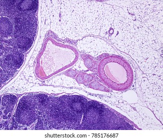


Vein Histology Images Stock Photos Vectors Shutterstock
9/3/15 Virtual Histology Main Navigation Help Skeletal muscle ls iron hematoxylin #46 Skeletal muscle ls #47 Skeletal Aorta Mallory stain #84 Aorta WeigertVan Gieson elastic stain #85 Aorta DMS106 Artery/vein/nerve # Aorta DMS107 Muscular artery Wiegart Stain DMS108 Vena cava DMS110 Vena cava Trichrome stainPublic Collections StLouisU Dr Smiths Virtual Histology Slides 001 Artery, vein, nerve, and skeletal musclesvsStart studying Artery, Vein & Nerve Histology Learn vocabulary, terms, and more with flashcards, games, and other study tools


Peripheral Nerve Histology Histology Flashcards Draw It To Know It


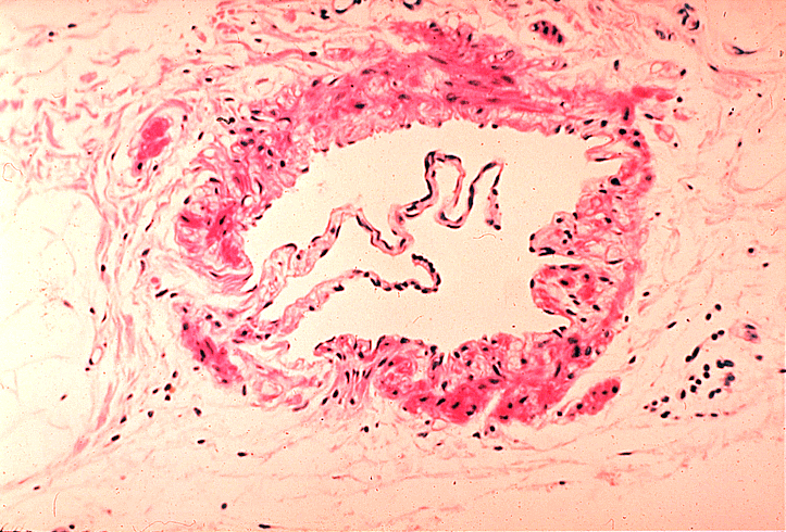
Small Veins And Muscular Arteries
Artery capillary cardiac muscle endo, myo, and epicardium elastic artery internal and external elastic laminae Purkinje fiber tunica intima, media, and adventitia vas vasi (pl vasa vasorum) vein venule Slide #170 (Artery, vein, and nerve) ThereArtery, Vein & Nerve 400x Arteries and veins are located throughout the body connecting to the heart They are part of the circulatory system that carries blood to and from the heart and other tissues and organsStart studying Histology Artery, Vein and Nerve Learn vocabulary, terms, and more with flashcards, games, and other study tools
:background_color(FFFFFF):format(jpeg)/images/library/3608/jvqXu9FdCLcYYymIaZRwA_Sinusoidal_capillary.png)


Blood Vessels Histology And Clinical Aspects Kenhub


Blood Vessels Lab
Basic Histology Artery and Vein You will often be able to distinguish arteries and veins, especally when they run together, as they usually do Arteries have thicker walls and tend to have narrower lumens They have to constrict and dilate to control how much blood flows where, and they must bear the powerful force generated by the heart Histology of blood vessels 1 Blood Vessels 2 Objectives • Introduction • Components of circulatory system • Blood vessels – Basic structure • Arteries Elastic & Muscular • Arterioles, Metaarteriole • Capillaries Continuous, Fenestrated, Sinusoidal • Venules, Veins • Clinical Correlation 3HG2121T Artery Vein Nerve Trichrome Stain Prepared Microscope Slide Artery, vein & nerve;
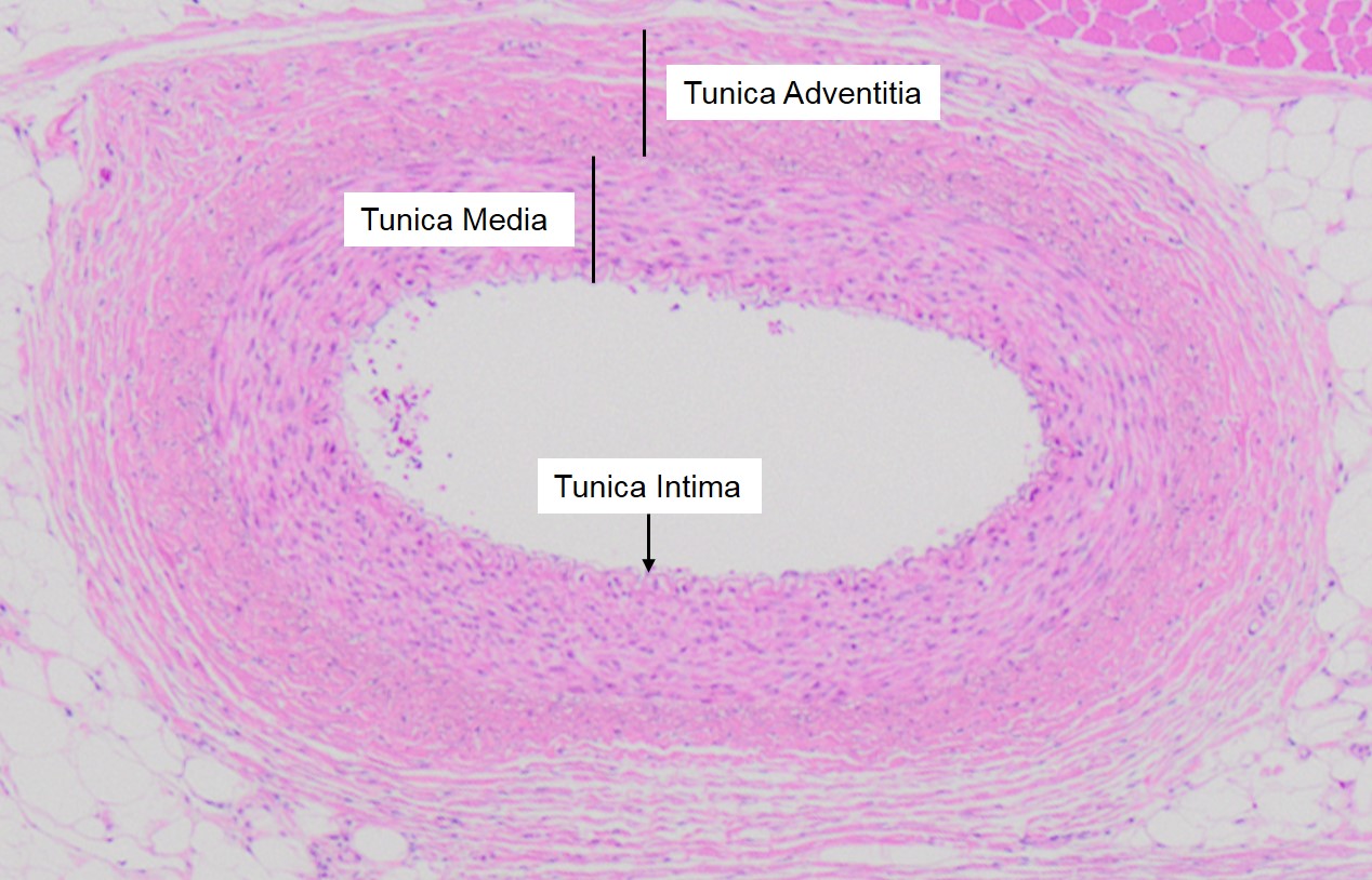


Vascular Tunics Veterinary Histology


Q Tbn And9gcri71nyfrsxcqs3 Hm7ddwfdy9idw7owspjrokzj W3pdqlfx4i Usqp Cau
Artery, Vein and Nerve $ 345 Artery, Vein and Nerve quantity Add to cart SKU PSH087 Category Histology Slides Additional information Additional information Weight lbs Dimensions125 × 1 × 3 in Related products View Cart Add to cart / Details CerebellumA slide of the Artery/Vein for Histology About Press Copyright Contact us Creators Advertise Developers Terms Privacy Policy & Safety How works Test new features © 21 Google


Histology Slides 1


Animal Organs Cardiovascular System Atlas Of Plant And Animal Histology


Artery Wikipedia


Artery Vein And Nerve Histological Slide Youtube


Histology Of Blood Vessels


Biology 12 Gladstone March 16


The Cardiovascular System Ross And Wilson Anatomy And Physiology In Health And Illness 11e


1


Artery Vein And Nerve Histology A P1 Flashcards Quizlet


Histology Slide Download Magscope Com Magscope Slide Bank Artery And Vein Magscope Teacher Resources Histology Slide Images To Save An Image Onto Your Computer Right Click It And Select Save As All Images Are Copyright Magscope They Are


Artery Vein Nerve Histology Diagram Quizlet
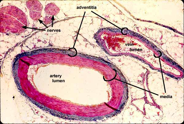


Siu Som Histology Intro
/GettyImages-464612781-edc0c4a8ab20491580bc3dd7acd4f4d2.jpg)


Capillary Structure And Function In The Body
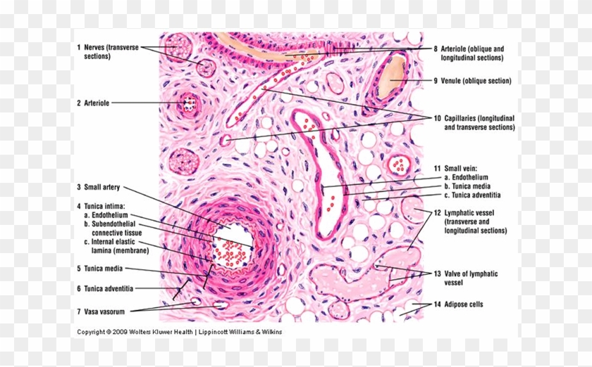


Blood Vessels Veins Arteries Capillaries Types Of Blood Vessels Histology Clipart Pikpng
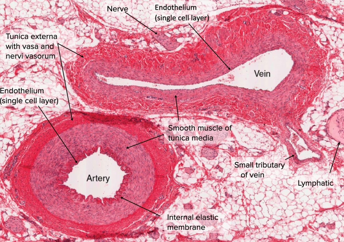


Arteries Concise Medical Knowledge
:background_color(FFFFFF):format(jpeg)/images/article/en/histology-of-the-vascular-network/qTcfa6edhRn9fZVNzTw_aCakVCMk7M8xtaJDDC6A4w_Artery.png)


Blood Vessels Histology And Clinical Aspects Kenhub


Arteries


Cardiovascular System Histology


Arteries
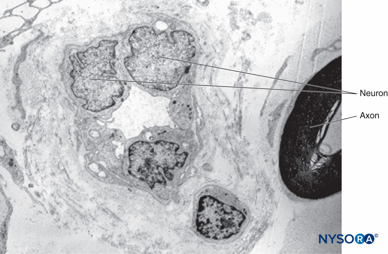


Connective Tissues Of Peripheral Nerves Nysora


Histology Laboratory Manual
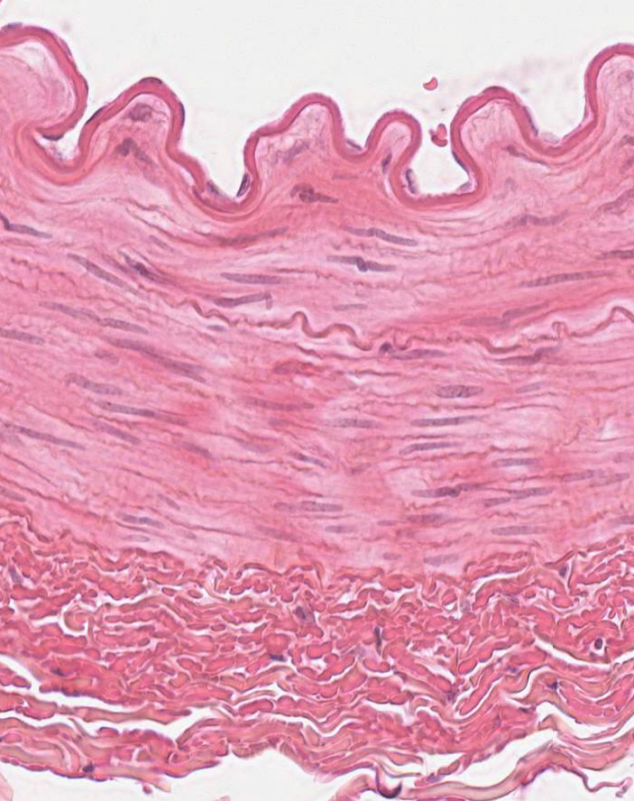


The Aorta


Artery Vein Nerve Prepared Microscope Slide 75x25mm Eisco Labs
:background_color(FFFFFF):format(jpeg)/images/library/3605/qqhmVxIKd7pxeygeKSM3xw_Muscular_artery_-_Coronary_artery__1_.png)


Blood Vessels Histology And Clinical Aspects Kenhub
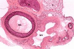


Cp Histology Anatomy Gu Som Flashcards Cram Com


Histology Slides Blood Vessels And Nerves Flashcards Quizlet
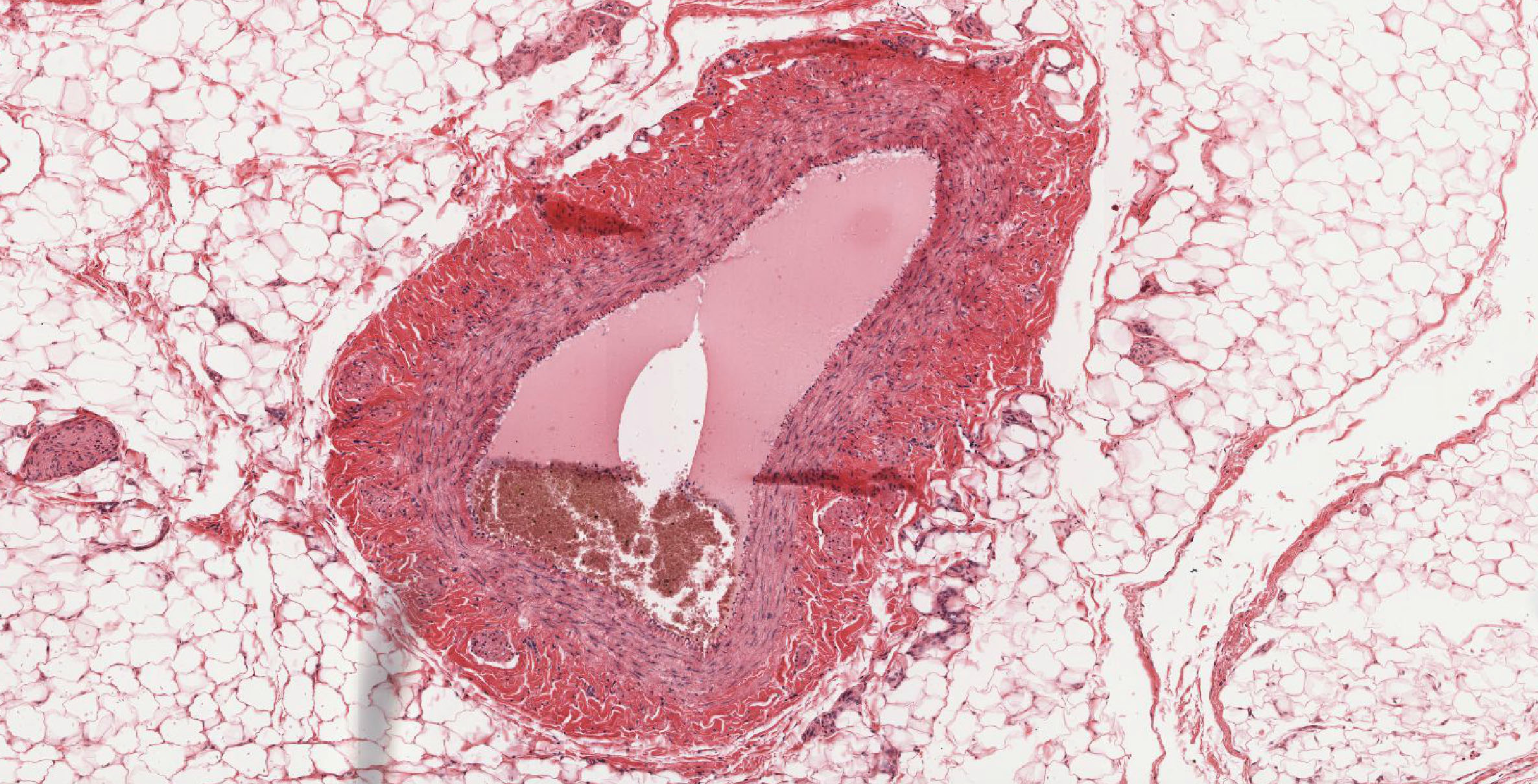


Cardiovascular System Histology


Blood Vessels Lab


Artery And Vein Histology Osmosis


Penis Histology Osmosis


Ultrastructure Of Blood Vessels Arteries Veins Teachmeanatomy


Van 4x Np Histology
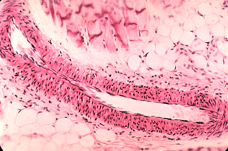


Small Veins And Muscular Arteries
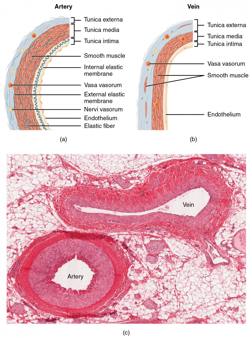


Structure And Function Of Blood Vessels Anatomy And Physiology Ii


Mammal Artery And Vein Transverse Section 32x Mammals Mammals Circulatory System Other Systems Comparative Anatomy Of Vertebrates Animal Histology Photos


Blood Vessels Lab


Mammal Artery And Vein Transverse Section 32x Mammals Mammals Circulatory System Other Systems Comparative Anatomy Of Vertebrates Animal Histology Photos


Photomicrograph Of A Vascular Bundle Of Tissue Including An Artery Vein And Several Nerves Human Tissue Human Anatomy And Physiology Autonomic Nervous System


Http Www Phlebology Org Wp Content Uploads 15 03 01 Anatomy Embryology Histology Macroscopic Anatomy Embryology Kalva Pdf


Untitled Document


Pin By Andres Sanchez On Histology Anatomy And Physiology Histology Slides Anatomy


Histology Vessels


3


Massasoit Instructure Com Courses Files Download Wrap 1
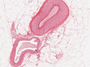


Cardiovascular System Histology
:background_color(FFFFFF):format(jpeg)/images/library/3607/bxosDBcdwLdq47dXkSy4A_Fenestrated_capillaries.png)


Blood Vessels Histology And Clinical Aspects Kenhub


Artery And Vein
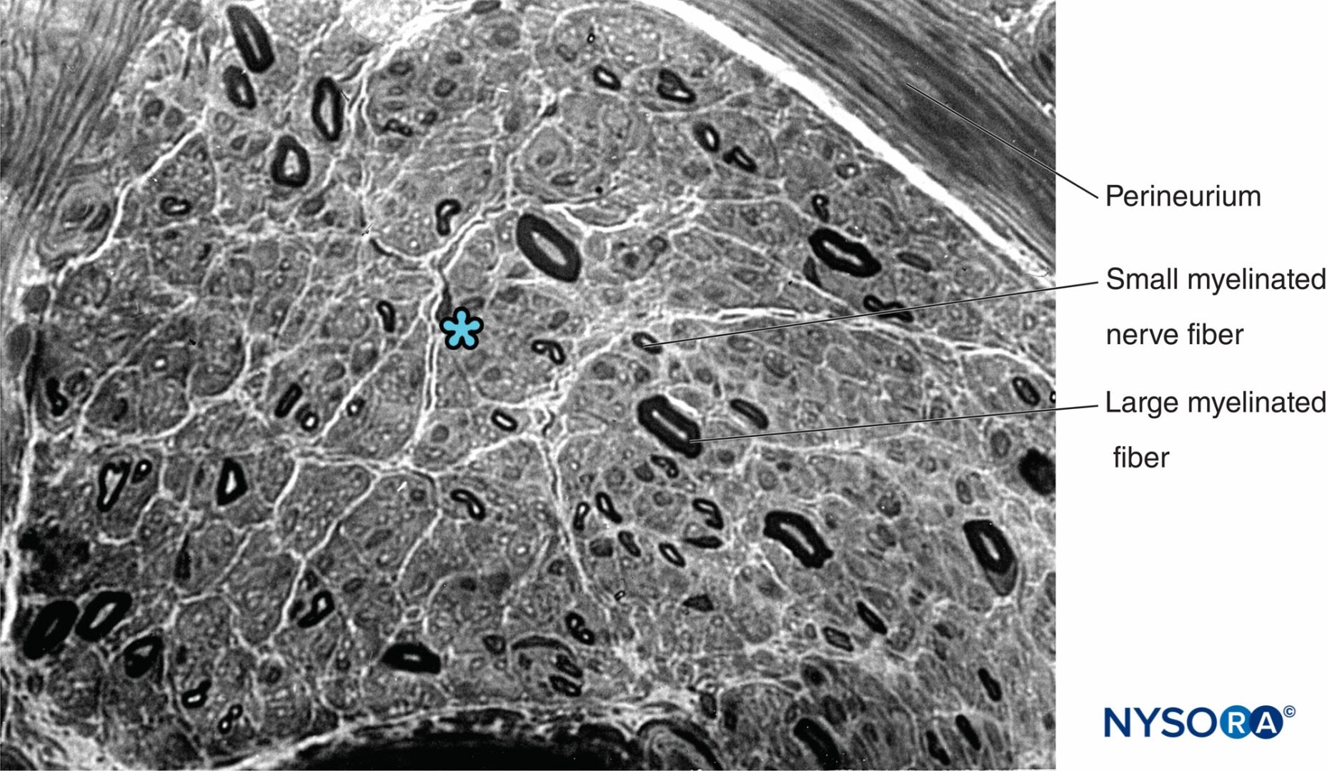


Histology Of The Peripheral Nerves And Light Microscopy Nysora


A Histology Of Common Hepatic Artery Day 90 Movat S Pentachrome Download Scientific Diagram
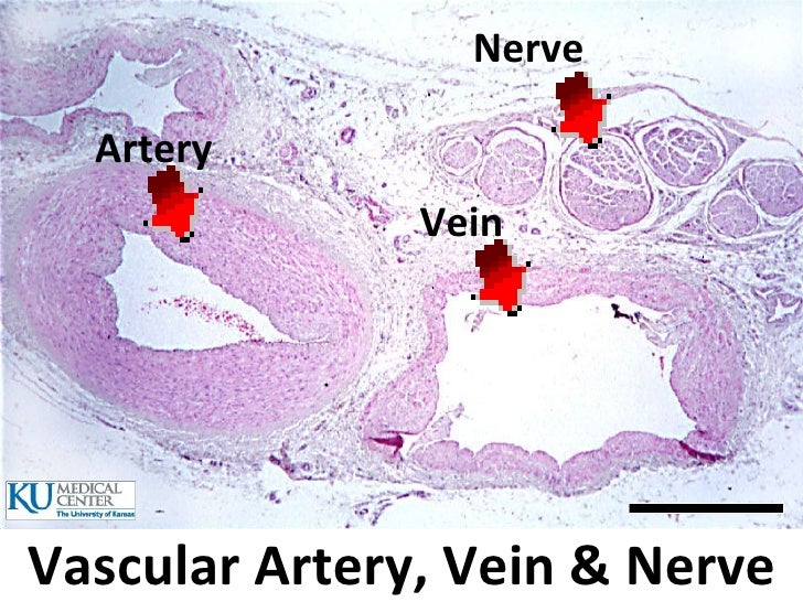


Histology Lab Image Information


Pin On Medical Laboratory Science


Untitled Document


Small Veins And Muscular Arteries


Nerve Structure Anatomy


Blood Vessels Knowledge Amboss


Pin On What I M Learning


Histology Of The Peripheral Nerves And Light Microscopy Nysora


Blood Vessel Color Images
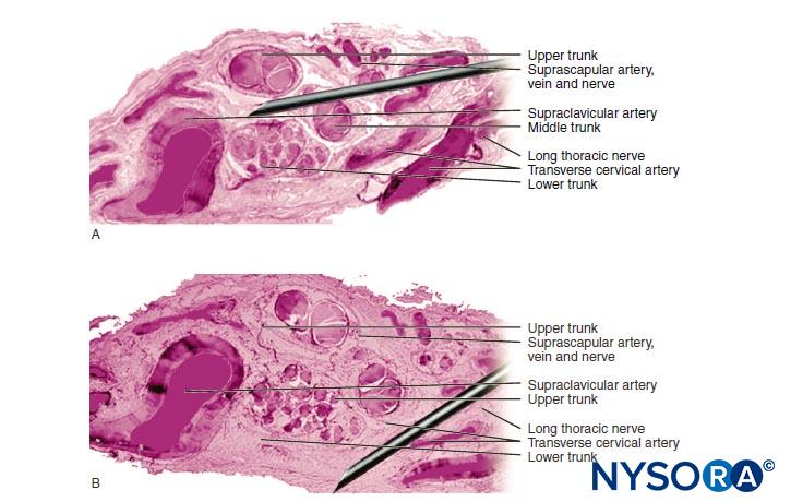


Complications And Prevention Of Neurologic Injury With Peripheral Nerve Blocks Nysora


Peripheral Nerves Definition Distribution Preview Histology Kenhub Youtube
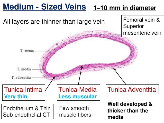


Histology Of Blood Vessels
:background_color(FFFFFF):format(jpeg)/images/library/3606/TIdQTQlRbloxtCUKcs2g_Arterioles.png)


Blood Vessels Histology And Clinical Aspects Kenhub


Histology Of Blood Vessels


Q Tbn And9gcqpuhblbr0wdzhvvdglabclwj5mimm Sdhcwm3qpjbwmc0u4fc Usqp Cau


Histology Slides 1


Histology Of The Retromolar Canal A Artery With A Thick Wall Download Scientific Diagram


Untitled Document


Arteries


Histology Slide Download Magscope Com Magscope Slide Bank Artery And Vein Magscope Teacher Resources Histology Slide Images To Save An Image Onto Your Computer Right Click It And Select Save As All Images Are Copyright Magscope They Are


Jerad Gardner Md Blood Vessels Nerves Usually Travel Alongside One Another This Section Of Normal Subcutis Shows A Nerve Vessel Weaving In And Out Of The Tissue Plane
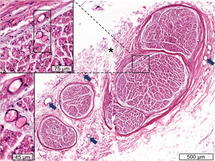


Cervical Vagus Nerve Morphometry And Vascularity In The Context Of Nerve Stimulation A Cadaveric Study Scientific Reports


Histolab3a Htm


Cardiovascular
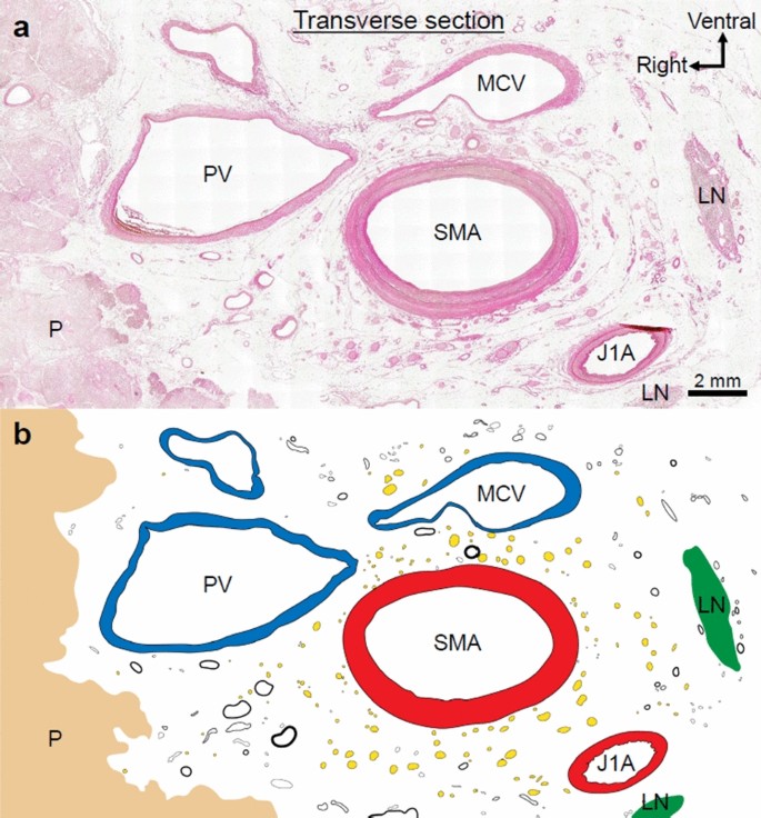


What Comprises The Plate Like Structure Between The Pancreatic Head And The Celiac Trunk And Superior Mesenteric Artery A Proposal For The Term P A Ligament Based On Anatomical Findings Springerlink


Untitled Document


Histology Laboratory Manual


Human Artery Vein Nerve Cross Section Prepared Microscope Slide Eisco Labs
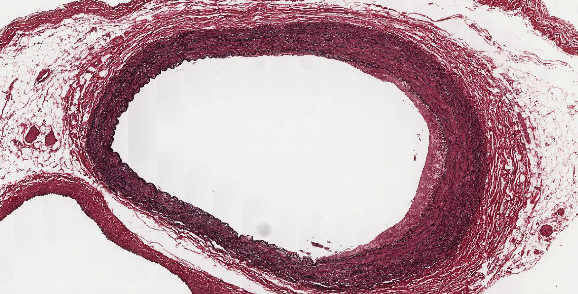


Cardiovascular System Histology


0 件のコメント:
コメントを投稿